- Home
- Microscope Pictures
Microscope Pictures of Hidden Lifeforms
These microscope pictures were taken by me with three different types of microscopes. The microscope cameras were connected to a laptop by a USB cable.
I am an amateur microscopist, so I don’t yet have the scientific names for some of the microscopic images shown, particularly the microscope pictures of pond water.
How I Took These Microscope Pictures
For making microscope images in my home lab, I use three types of microscopes. The first is a pocket microscope. The second is a handheld USB digital microscope. The third is a compound microscope with a camera attachment.
The pocket microscope is the Carson Microflip 100-250x with a cell
phone attachment. I purchased the bundle with 2 sets of prepared slides,
under $30 for all.
The USB microscope is an AmScope 50x to 500x digital microscope. It came with a stand, and attaches to the USB port on a computer. There is no eyepiece, so the only way to view specimens is through the computer.
The third microscope is a Swift compound microscope, model SW380T. This is a compound trinocular microscope with 40x to 2500x magnification. It came with a mechanical stage, and a 5MP camera.
For the latter two, I connected the microscope to a MacBook laptop and used the Photo Booth app to capture photos and videos.
Studying Pond Water Organisms with a Microscope
Of all the specimens I’ve examined under the microscope in
my home lab so far, pond water is the most interesting to me. You can use these specimen images for kids to get them interested in science.
Some of the lifeforms that can be seen in a pond water sample include bacteria, protozoa, algae, rotifers, ciliates, diatoms, cyanobacteria, water fleas called Daphnia, hydra, nematodes, and water mites.
Detailed Microscope Images of Pond Organisms
Visit the YouTube link to see them all in action in the video.
 Diatoms
 Cyanobacteria, algae
 Diatoms, algae, and others
 Diatoms
See the diatoms in action in the Pond Water Under Microscope video on my YouTube channel:
Under the Microscope: Human Hair Examination
Under the microscope you can study the condition of the hair shaft, cuticle layers, and medulla.
If a hairbrush is used by everyone in the family, you'll see an assortment of colors and conditions. Notice that some of the hairs in this sample are transparent. Those came from a gray-haired person.
It is possible to distinguish animal hair from human hair by examining the cuticle patterns.
Fiber examination can tell us whether the specimen is natural (plant or animal) or synthetic.
The first two hair pictures were taken with the Amscope digital USB microscope. The others were taken with the Swift compound microscope.
 Various hairs from a hair brush
 Various hairs, same sample
 Human hair cuticle
 Human hair 40x objective (400x)
 Dog hair cuticle
 Synthetic carpet fibers - no cuticle patterns
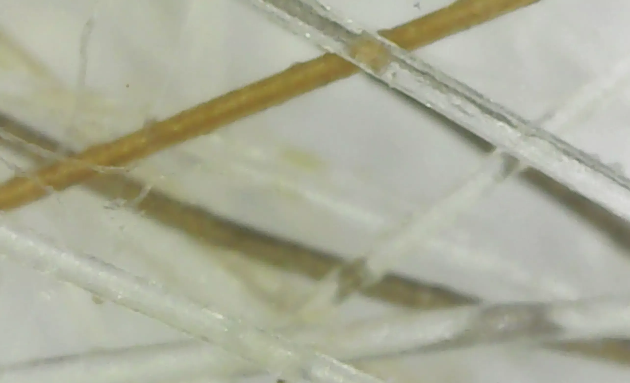
Examining Various Fibers Under the Microscope
In dryer lint, along with fibers, you might also find hair. In vacuum cleaner dirt you will find fibers from carpet, clothing, hair, skin, and soil.
Crime scene evidence is sometimes gathered by vacuuming to collect fibers and hair. Individual fibers can then be compared to reference samples. For example, evidence collected from a suspect might match evidence collected from a crime scene.
Some clothing is made from animal fibers, such as cashmere, wool from sheep and alpacas. You can tell if a fiber is from an animal by looking for a cuticle pattern.
Other clothing fibers might be natural, like cotton and bamboo, others might be nylon, rayon, or polyester.
 fibers in dryer lint
 fibers collected by vacuum cleaner
 Wool from blue scarf
 Wool (sheep fibers)
Prepared Slides and Other Microscope Pictures
These images were taken on a cell phone attached to a pocket microscope.
Prepared slides come in sets, and may be from insects, tissue samples, or plants. The first two below, are from an insect set.
The next two pictures are examples of what might be used by coin collectors to grade coins.
 Insect wing picture from prepared slide
 Bee part from prepared slide
 The number 8 on a coin
 The letter G on a coin
About The Microscope Pictures
I collected all the samples from things around my home. The pond water is from a small pond in my backyard, next to a 110 year old spring house. The hair is from me and my dog, Remi. The fibers are from my dryer, vacuum cleaner, wool scarf and bedroom carpet.
Some samples, the insects for example, came from prepared slides I purchased along with the Carson microscope.
I used the Carson Microflip and cell phone attachment to take pictures with my iPhone. Most of the photos taken from this microscope will be in a round format.
I used the AmScope 50x to 500x digital microscope for some of the hair and vacuum cleaner samples.
My Swift compound microscope camera provided the higher magnification pictures of pond water, hair, and textile fibers. I captured the photos and videos on my MacBook with the Photo Booth app.
- Home
- Microscope Pictures
You might like these
How Does a Microscope Work?
How Does a Microscope Work? Read this to learn the answer, as well as learn the parts of a microscope.
Jobs That Use Microscopes
What are jobs that use microscopes? Did you think of them all? Check out list.
The Marvelous Mechanical Stage
The mechanical stage of a microscope allows you to smoothly and precisely move your slide while viewing. Discover how they work and other types of stages.


If you visit an
affiliate from a link (clearly marked) on my site, I may receive a small commission at no cost
to you. As an Amazon Associate I earn from qualifying purchases. As an eBay Partner, I may be compensated if you make a
purchase through a link.
This search feature contains ads at the top and search results below that. Just scroll down to see the pages that match your search.
______________________
Recent Articles
-
Microscope Activities for Middle School Students
 Microscope activities for middle school students for engaging lessons. They work for home schoolers and middle school students in traditional classrooms.
 -
Microscope Pictures
 Microscope pictures of pond life, hair and fiber samples, and yogurt taken by a digital USB microscope and a compound microscope camera connected to a computer.
 -
How to Draw a Microscope
 If you've been given this assignment for science class, here's how to draw a microscope, step-by-step, with microscope pictures.