How Does a Microscope Work?
The world around us is full of intricate details that often go unnoticed due to the limitations of the human eye. Microscopes, with their unique ability to magnify objects, help bridge this gap in our perception and broaden our understanding of the universe, small scale to large. But how does a microscope work? That’s a question we’ll tackle in this article, diving deep into the world of microscopes.

A microscope’s primary function is to magnify small objects so they can be seen clearly with the naked eye. It does this by bending or refracting light that passes through a specimen, producing an image that appears much larger than the object’s actual size.
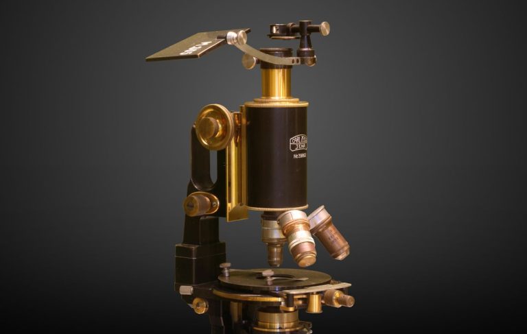
Before understanding how a microscope works, it is helpful to understand the parts of a microscope. The microscope is made up of several components, each playing a unique role in the image projection process.
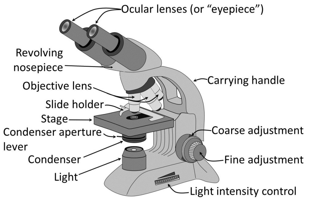
At the top of the microscope, we find the eyepiece or ocular lens. This part of the microscope is where we place our eye to observe the magnified image. The eyepiece has its own magnification power, typically 10x.
Connecting the eyepiece to the base of the microscope is the body tube, an integral structural component. Most importantly, it houses the prism that redirects light up through the eyepiece.
Another crucial part of the microscope is the objective lens. Located at the other end of the body tube, the objective lens is much closer to the specimen being observed. There are sometimes three or four objective lenses on a rotating turret, each providing different magnification levels, ranging from 4x to 100x.
The microscope stage, located below the objective lenses, is where you place the specimen or slide being observed. The stage holds the specimen in place and some, called a mechanical stage, can be moved up and down to bring the sample closer to or further from the objective lens.
Illumination is key in any optical instrument. The light source or illuminator, located below the stage, is there to shed light on the specimen. This not only allows us to see the specimen but also forms the image that is then magnified by the lenses.
Summary of main parts of a microscope:
- The eyepiece and ocular lens at the top of the microscope where we place our eye to observe the magnified image
- The body tube, connecting the eyepiece to the base of the microscope, houses the prism that redirects light up through the eyepiece
- The objective lens, located at the other end of the body tube, near the specimen being observed and offering different magnification levels
- The stage, where the specimen or slide being observed is placed
- The light source or illuminator, located below the stage to shed light on the specimen
Yes, but How Does a Microscope Work?
Now, how do microscopes work?
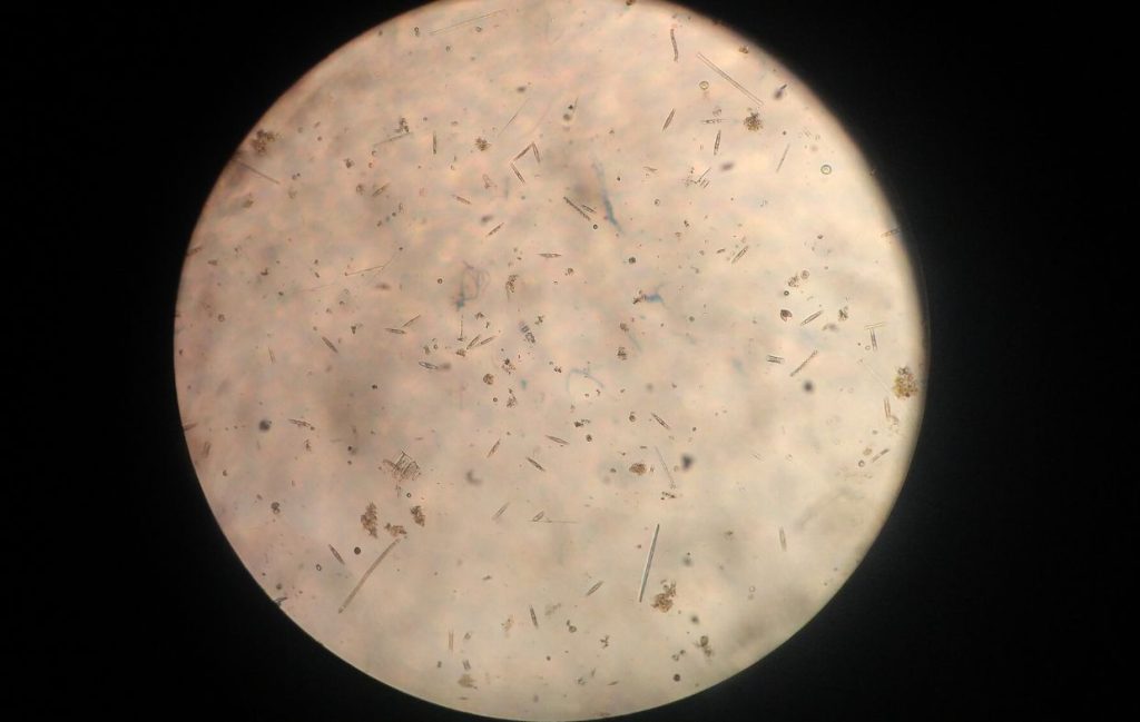
In a compound microscope, light emissions from the illuminator pass through the specimen on the stage. This light then goes through the objective lens, which magnifies the image and sends it up through the body tube and the ocular lens for you to view.
In a microscope, the prism is used to redirect light. It is housed inside the body tube and it collects the light passing through the specimen and objective lens, then redirects it upwards to the eyepiece or ocular lens, enabling the user to view the magnified image.
Microscope magnification is achieved by multiplying the magnification of the objective lens by the eyepiece lens. For example, if you’re using a 10x eyepiece and a 40x objective, you will get an overall magnification of 400 times.
Microscopes are not just about magnification; they also resolve the image. Resolution is the microscope’s ability to distinguish two points on a specimen as separate entities, ultimately determining the clarity of the image. We commonly think of this as focus.
Apart from understanding the parts and workings of a microscope, it is also necessary to maintain it properly and carefully. Microscopes are precision instruments, and even a small amount of dust or debris can affect their functioning and the image quality they can produce.
To clean the lenses of a microscope, use only lens-specific cleaning materials. Rough materials or home cleaning products might cause scratches or leave films that would affect the quality of the image.
Make sure the microscope is covered when not in use to protect it from dust and accidental damage. It’s also a good practice to store it in a cool, dry place away from direct sunlight.
Annual professional servicing is a good idea if your microscope gets heavy use. This will involve cleaning, lubrication, and alignment checks, among other steps.
Understanding the structure and functionality of a microscope is not just intriguing, but it also helps users maintain the instrument and troubleshoot basic issues.
While modern technology has introduced more complex types of microscopes, like scanning electron microscopes or atomic force microscopes, the essential principle remains the same – magnify the small things to help us better understand the microscopic world around us.
The microscope has undeniably been one of the most critical tools in the journey of scientific discovery. Its integration in countless fields, such as biology, medicine, geology, and even art, reaffirms its significance.
In sum, understanding how a microscope works is not only fascinating but also contributes to a greater understanding and appreciation of the significant impact this tool has made on our world. Whether you’re a student grappling with your first lab assignments or a professional scientist exploring the frontiers of your field, the microscope is indeed a key to a universe unseen.