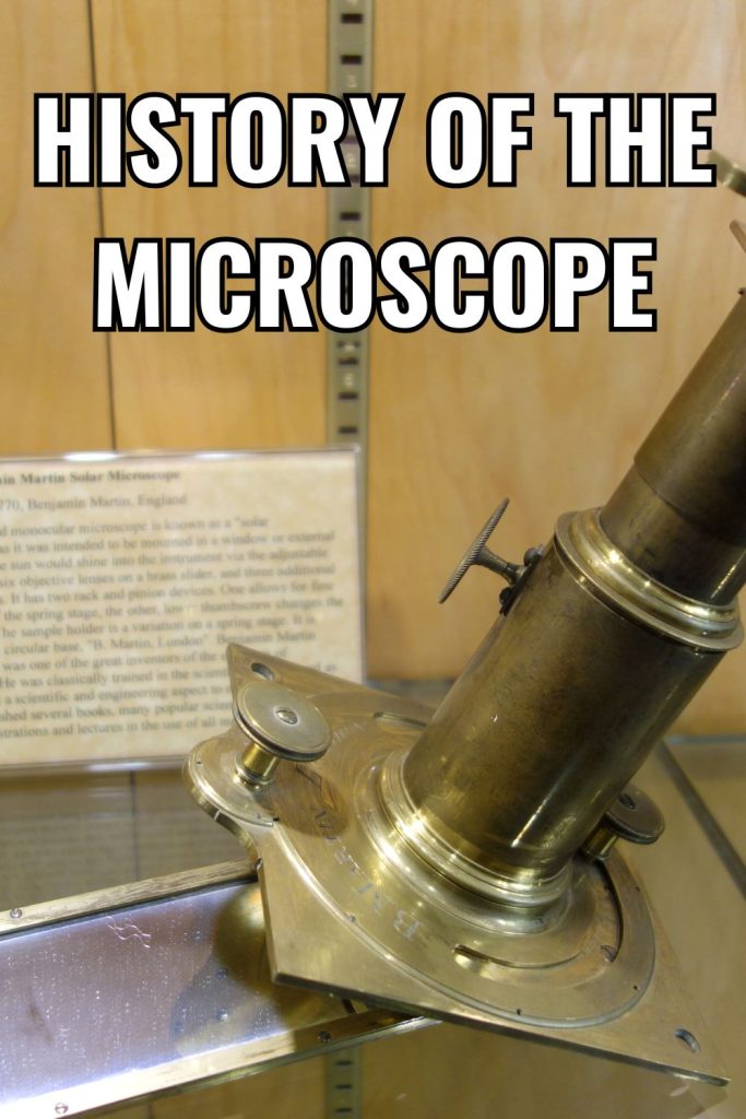
The Intriguing History of the Microscope
The history of the microscope could fill a large book, or more. Here, we will highlight significant points in the history of microscopes.
This incredible instrument has allowed us to peer into the tiniest corners of the natural world and make groundbreaking scientific discoveries. But the story of the microscope’s invention is a fascinating tale that spans centuries. Let’s explore the history of the microscope and find out who we have to thank for this remarkable invention.

Microscopy in Ancient Times
The earliest origins of the microscope actually date all the way back to ancient times. As far back as the 1st century AD, the Romans were using glass to make simple magnifying lenses.
These early lenses were used for all sorts of things like reading, examining gemstones, and even cauterizing wounds.
But it would be many centuries before these primitive lenses evolved into the first true microscopes.
16th Century Simple Microscopes
Fast forward to the late 16th century and we have the first microscope inventors on record – Dutch spectacle makers Zacharias Janssen and his father Hans. Around 1590, the Janssens experimented with placing multiple lenses in a tube and discovered that nearby objects appeared greatly enlarged. These early compound microscopes were more of a novelty than a scientific tool, with images that were blurry and distorted. But the Janssens had planted the seeds for the microscope’s evolution.
17th Century Microscope Inventors
The next major milestone in the history of the microscope came from the famous Italian astronomer Galileo Galilei. In 1609, he developed a compound microscope that had a convex and a concave lens. This provided 3x magnification and produced much clearer and brighter images than the Janssen design. Galileo’s microscope was one of the first to actually be used for scientific purposes, like observing insect eyes and bee stingers.
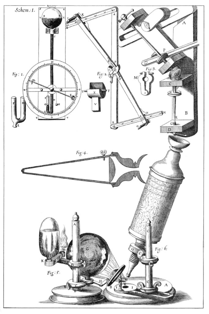
But the microscope was about to get a major upgrade. In 1665, English physicist Robert Hooke created a microscope with a groundbreaking new design. His “Hooke microscope” used three lenses and a stage light, which allowed for magnification up to 50x. Hooke used it to make the first detailed observations of plant cells and insects. He even coined the term “cell” after noting how plant cells looked like the small rooms that monks lived in. Hooke’s beautifully illustrated book “Micrographia” introduced the unseen microscopic world to the public for the first time.
See the book at Project Gutenberg (Micrographia).
While Hooke was making his observations in England, over in Holland, a draper named Antonie van Leeuwenhoek was quietly pioneering the single-lens microscope. Leeuwenhoek’s microscopes were tiny – about the size of a large pen – but they were marvels of engineering. His precise grinding techniques and use of a single spherical lens produced microscopes with magnifications of over 200x with surprisingly clear images.
He discovered a whole world of microscopic life that no one even knew existed. He was also the first to see red blood cells and sperm cells.
Leeuwenhoek’s discoveries marked the birth of microbiology and cell biology. For this, he is often called the “Father of Microbiology”.
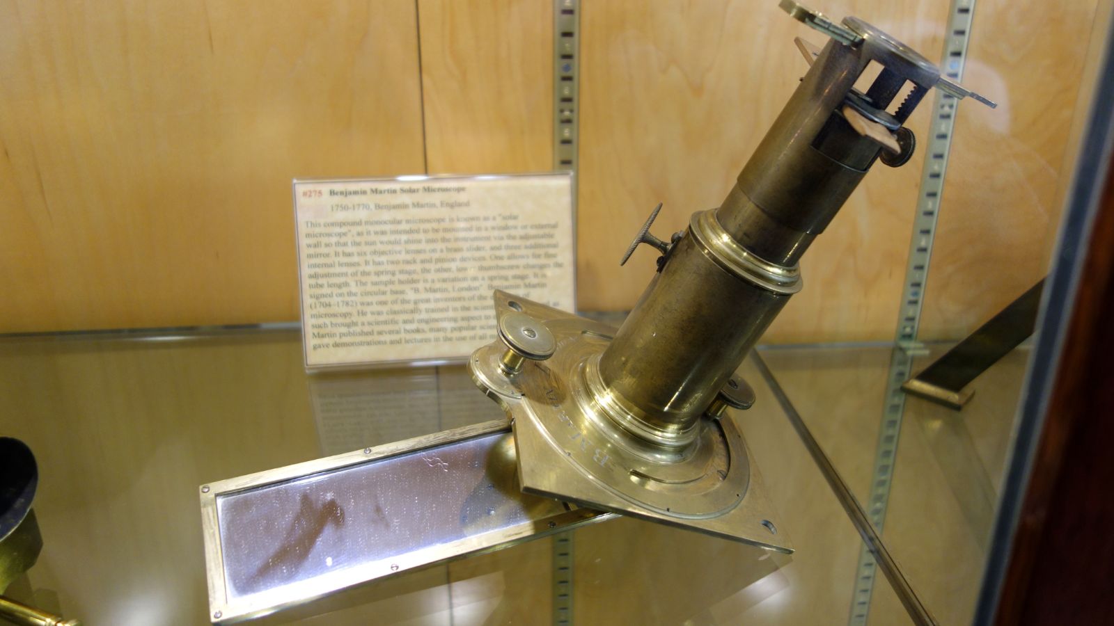
18th – 19th Century Evolution of Microscope Technology
Throughout the 18th century, the microscope continued to evolve with new technical innovations. Improved lenses and ways to correct optical aberrations led to microscopes with higher magnification and better image quality.
By the early 1800s, microscope technology had advanced to the point where compound microscopes were capable of 1000x magnification – enough to see most bacteria. These microscopes became essential tools for scientists studying everything from microorganisms to human cells and tissues.
Lister, Zeiss, and Abbe: Key Figures in the History of the Microscope
The mid-1800s brought a golden age for microscopy, with several major advancements in quick succession. In the 1850s, Carl Zeiss, a German manufacturer of optical equipment, began producing microscopes with scientifically designed lenses that were much more precise than previous models. Together with the physicist Ernst Abbe, Zeiss pioneered new ways to improve the resolution and magnification of microscopes.
One of their key inventions was the oil immersion lens, where the objective lens is immersed in a drop of oil that has the same refractive index as glass. This allowed for much higher magnifications by reducing the refraction of light as it passed through the lens. With these new lenses, microscopes could finally provide detailed views of the interior structures of cells.
In 1869, Ernst Abbe published a mathematical theory that would become the foundation of modern microscope design. Abbe’s Diffraction Theory described the physical limitations on the resolution of microscopes. He proposed that microscopes had a finite resolution that was limited by the wavelength of light and the aperture of the lenses. Abbe’s theory guided the design of microscope lenses for improved resolution and correction of optical aberrations.
The late 19th century brought the development of specialized microscopes for different applications. The petrographic microscope, for example, was developed for observing rock and mineral samples using polarized light. The metallurgical microscope was used for examining the microstructure of metals and alloys. And the comparison microscope allowed forensic scientists to compare bullets, tool marks, and documents side-by-side.
Microscopes in the 20th Century
The 20th century introduced new ways of imaging that went beyond the abilities of light microscopes. In 1931, German engineers Ernst Ruska and Max Knoll built the first electron microscope, which used a beam of electrons instead of light to view specimens. Electron microscopes took magnification to a whole new level, with some able to magnify objects up to 10 million times. This allowed scientists to finally see the details of viruses, molecules, and even individual atoms.
Other types of microscopes also emerged in the 20th century. The phase contrast microscope, invented in 1934, allowed scientists to view transparent living cells by making subtle changes in light phase visible. The scanning tunneling microscope, developed in 1981, could image individual atoms on the surfaces of materials. And fluorescence microscopes, which use glowing molecular labels, opened up new ways of imaging living cells and the localization of proteins.
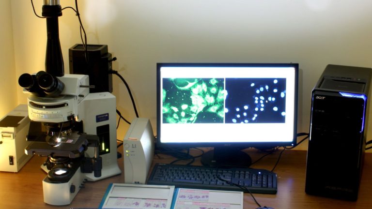
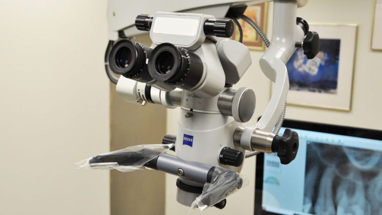
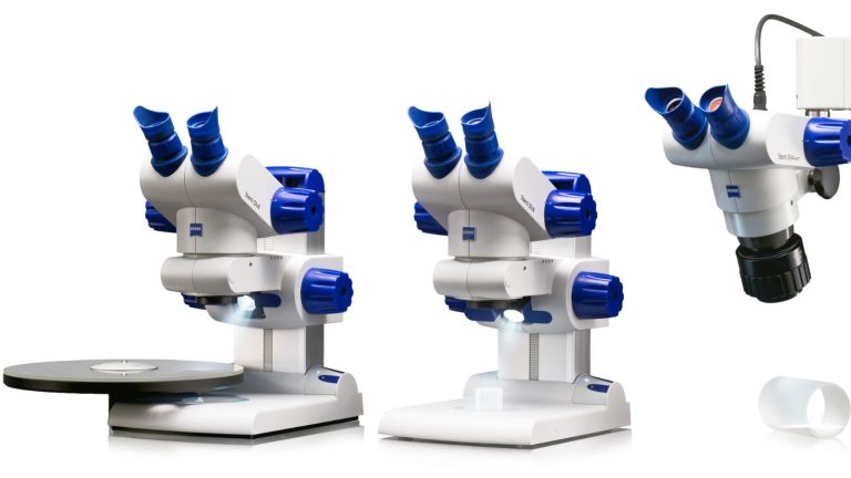
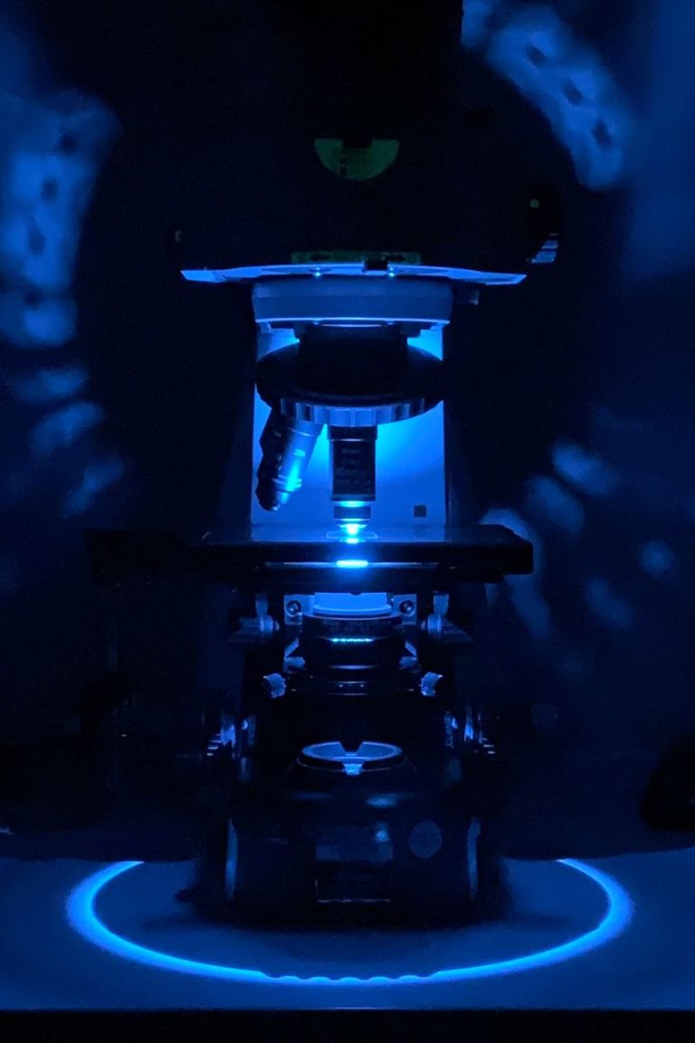
Modern Microscopes
In the 21st century, microscopy has become a high-tech field that combines cutting-edge optics, digital sensors, and advanced software. New techniques like super-resolution microscopy allow us to take images with resolutions below the diffraction limit of light. Microscopes with built-in spectrometers can identify the chemical makeup of samples. And automated microscopes driven by artificial intelligence can quickly scan and analyze thousands of samples.
Learn more about microscopes today:
In Conclusion on the History of Microscopes
From the first glimpses of the invisible world to the latest advances in nanotechnology, the history of the microscope spans over 400 years of scientific discovery and innovation. So many brilliant minds have contributed to the invention and evolution of this groundbreaking scientific tool – Zacharias and Hans Janssen, Galileo Galilei, Robert Hooke, Antonie van Leeuwenhoek, Joseph Jackson Lister, Carl Zeiss, Ernst Abbe, and many more.
So who invented the microscope? There’s no single answer.
Instead, the microscope was born out of the collective ingenuity of inventors and scientists over centuries – each building on the work of those who came before to bring us the incredibly powerful micro-imaging tools we have today. As we continue to push the boundaries of microscopy, who knows what new wonders it will reveal? The story of the microscope is still being written, and the next groundbreaking innovation could be just around the corner.